Diagnosing Plasma by Optical probing
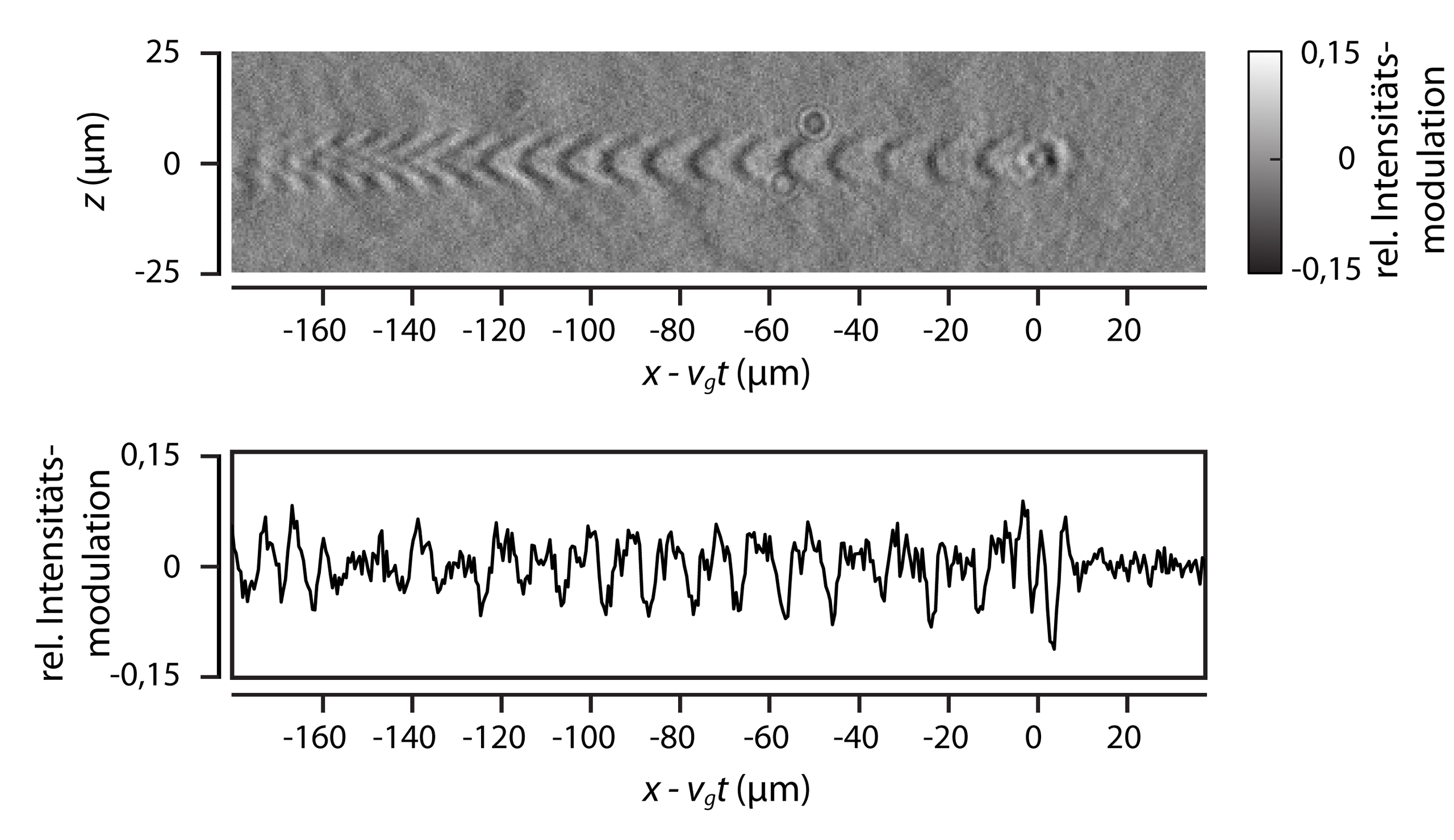
The direct investigation of the laser-plasma interactions in Laser Wakefield Acceleration (LWFA) is especially challenging considering that the plasma wave’s micrometer-scale size and its femtosecond-scale temporal evolution must be resolved simultaneously. The interaction region can be recorded by illuminating it with a second laser pulse, typically called a “probe pulse” or simply “probe”, and imaging the region onto a sensor. In order to record fine details of the interaction the imaging system used must achieve the requisite spatial resolution and the probe pulse used as illumination must have a pulse duration significantly shorter than that of the driving laser pulse. For exactly this purpose, members of the IOQ and HI-Jena have developed an imaging technique named “Few-Cycle Microscopy (FCM)”. This technique uses a fraction of the driving pulse’s energy to be the probe pulse. The probe is spectrally broadened via a nonlinear process, and then temporally compressed to a pulse duration of a few femtoseconds, i.e., a few optical cycle pulse duration in the near-infrared. Combined with a microscope designed for the visible and near-infrared spectra, the FCM system can directly image the plasma wave that is crucial to the LWFA process. First implemented on the JETi40 laser system, the JETi200 laser now has its own FCM setup and with it the ability to image plasma waves and their evolution using shadowgraphic imaging, as well as to quantify the azimuthal magnetic fields in the plasma wave via polarimetric imaging. Furthermore, density measurements of the electron plasma distribution can also be performed using interferometric methods. Details of the laser-plasma interaction and plasma wave that were once only visible in numerical simulations can now be measured experimentally thanks to Few-Cycle Microscopy.

The achievable peak kinetic energy of the accelerated electron bunches is inversely proportional to the electron density in the plasma, meaning that the electron plasma density must be reduced to reach higher electron bunch energies within a single LWFA stage. In order to preserve a high image contrast in the shadowgraphic images, the central wavelength of the probe pulse must be shifted to longer wavelengths. This is possible with the use of an optical parametric amplifier. Once the central wavelength of the probe has been shifted, the probe once again undergoes spectral broadening and temporal compression to a pulse duration at the few-cycle level. The previously mentioned imaging schemes can then be used to provide detailed images and measurements of LWFA in this novel, low-density regime. The FCM diagnostic system not only lends itself to detailed measurements of laser-driven plasma accelerators, but can also be implemented at particle-driven plasma accelerators, such as in the FLASHForward project at DESY in Hamburg where electron bunches will be used to drive the plasma wave in Particle Wakefield Acceleration.