we are glad to provide you — after a fairly long break related to a complete renewal of our institute website — with a new issue of the newsletter of the Helmholtz Institute Jena. Below you find informations and news about recent activities of our institute.
Kind Regards,
Helmholtz Institute Jena
Structured Illumination Ptychography boosts Table-Top Nanoscale Imaging in the Extreme Ultraviolet
Fig. 1. a) Complex EUV image of a part of a microelectronic solid-state disc. b) Scattering quotient map allowing for identification of the material composition on the nanoscale [1].
Fig. 2. High-resolution EUV ptychography reconstruction of E. coli bacteria. For a single E. coli cell in the state of cell division, which is indicated by a pink box, the scattering quotient is calculated and shown in the inset. Low values of the scattering quotient correspond to the cell wall, which is dominated by carbon hydrates, while the large scattering values correspond to proteins, lipids, and other biological compositions [2].
Microscopy with extreme ultraviolet (EUV) radiation holds promise for high-resolution imaging with excellent material contrast, due to the short wavelength and numerous element-specific absorption edges available in this spectral range. At the same time, EUV radiation has significantly larger penetration depths than electrons.
By combining a high photon-flux, table-top EUV source, structured EUV illumination in a scanning lensless imaging setup, and advanced computational Ptychography algorithms, an important breakthrough has recently been achieved by researchers at the HI-Jena.
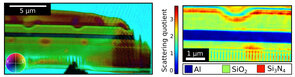
A record-high spatial resolution of 16 nm has been demonstrated in combination with highly-accurate amplitude- and phase-imaging over wide fields of view [1]. In a first demonstration, the complex transmission of an integrated circuit (part of a solid-state disc) was precisely reconstructed as shown in fig. 1 a). By calculating the scattering quotient as the ratio of the observed phase shift and attenuation in each pixel, the nanoscale material composition is identified, as shown in fig. 1 b).
So far, high-performance lensless imaging at short wavelengths was restricted to large-scale facilities, where beam time is valuable and scarce. The presented experiments utilizing laser-generated coherent EUV radiation (high order harmonics) demonstrate that such capabilities get now available in compact table-top setups, which can be implemented in standard research laboratories but also in industrial or clinical environments.
Potential applications are found in materials and life sciences and many other areas. For example, high-resolution studies of the composition and the nano-structure of compound battery materials or the investigation of sub-cellular structural and chemical changes caused by diseases or infections get feasible. The highly-accessible setups will facilitate in-house, rapid-cycle, and fast-feedback studies without the need for travel and beam times and will thus speed up future developments in many fields.
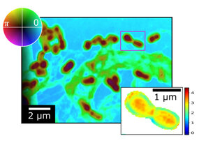
Encouraging results have already been obtained when imaging microorganisms in the novel XUV microscope [2]. Two dried model specimens: germlings of a fungus (Aspergillus nidulans), and bacteria (Escherichia coli) cells have been investigated with a half-period spatial resolution of 58 nm. Again a scattering quotient micrograph uncovers the composition of the sample averaged along the propagation direction in each pixel of the image. In both investigated samples, different biological compositions (such as proteins, lipids, and hydrocarbons) have been obtained and successfully assigned to the internal functional units of the respective microorganisms. Example images for E. coli bacteria are shown in fig. 2. This first demonstration underlines the potential of imaging in the extreme ultraviolet spectral region and opens up new opportunities for life-science applications.
Mit einem neuartigen Verfahren lassen sich Laserstrahlen berührungsfrei in der Luft ablenken. Das dabei eingesetzte unsichtbare optische Gitter aus Luft ist nicht nur…
Philip Pfäfflein wurde für sein Poster “Precision x-ray spectroscopy of He-like uranium employing metallic magnetic calorimeter detectors” mit einem Posterpreis der …
In einem kürzlich erschienenen Artikel zeigt ein internationales Forscherteam des Helmholtz-Instituts Jena, des GSI Helmholtzzentrums für Schwerionenforschung, der…
Das Helmholtz-Institut Jena unterstützt das diesjährige MINT-Festival in Jena, mit dem Schüler*innen aller Altersstufen für Mathematik, Informatik, Naturwissenschaften…
Ringleb S,
Kiffer M,
Stöhlker T,
Vogel M.
Collisional thermalisation behaviour of highly-charged-ion plasmas in a Penning trap.
Journal of physics / B.
2025 May;
58(9):095202 -.
[DOI][File]
Schmelz M,
Shvab V,
Peiselt K,
Kunert J,
Zakosarenko V,
Stöhlker T,
Oelsner G,
Stolz R.
Highly Sensitive DC SQUID Arrays for the Readout of Optical TES at mK Temperatures.
IEEE Transactions on Applied Superconductivity.
2025 May;
35(3):1 - 4.
[DOI][File]
Lenski M,
Xu Q,
Gierschke P,
Wang Z,
Heuermann T,
Jauregui C,
Limpert J.
Average-power scaling of in-band pumped, Tm-doped, fiber laser systems for high peak power pulsed operation.
Optics express.
2025 Apr.;
33(7):16029 -.
[DOI][File]
Dellmann SF,
Glorius J,
Litvinov YA,
Reifarth R,
Varga L,
Aliotta M,
Amjad F,
Blaum K,
Bott L,
Brandau C,
Brückner B,
Bruno CG,
Chen R-,
Davinson T,
Dickel T,
Dillmann I,
Dmytriev D,
Erbacher P,
Forstner O,
Freire-Fernández D,
Geissel H,
Göbel K,
Griffin CJ,
Grisenti RE,
Gumberidze A,
Haettner E,
Hagmann S,
Heftrich T,
Heil M,
Heß R,
Hillenbrand P,
Hornung C,
Joseph R,
Jurado B,
Kazanseva E,
Khasawneh K,
Knöbel R,
Kostyleva D,
Kozhuharov C,
Kulikov I,
Kuzminchuk N,
Kurtulgil D,
Langer C,
Leckenby G,
Lederer-Woods C,
Lestinsky M,
Litvinov S,
Löher B,
Lorentz BA,
Lorenz E,
Marsh J,
Menz E,
Morgenroth T,
Mukha I,
Petridis N,
Popp U,
Psaltis A,
Purushothaman S,
Rocco E,
Roy P,
Sanjari MS,
Scheidenberger C,
Sguazzin M,
Sidhu RS,
Spillmann U,
Steck M,
Stöhlker T,
Surzhykov A,
Swartz JA,
Tanaka Y,
Törnqvist H,
Vescovi D,
Volknandt M,
Weick H,
Weigand M,
Woods PJ,
Yamaguchi T,
Zhao J.
First Proton-Induced Cross Sections on a Stored Rare Ion Beam: Measurement of Te 118 ( p , γ ) for Explosive Nucleosynthesis.
Physical review letters.
2025 Apr.;
134(14):142701.
[DOI][File]
Gies H,
Picciau M.
Mean-field theory for self-interacting relativistic Luttinger fermions.
Physical review / D.
2025 Apr.;
111(8):085001.
[DOI][File]
Pfäfflein P,
Weber G,
Allgeier S,
Andelkovic Z,
Bernitt S,
Bondarev A,
Borovik A,
Duval L,
Fleischmann A,
Forstner O,
Friedrich M,
Glorius J,
Gumberidze A,
Hahn C,
Herfurth F,
Hengstler D,
Herdrich MO,
Hillenbrand P,
Kalinin A,
Kiffer M,
Kroeger FM,
Kubullek M,
Kuntz P,
Lestinsky M,
Litvinov YA,
Löher B,
Menz EB,
Over T,
Petridis N,
Ringleb S,
Sidhu RS,
Spillmann U,
Trotsenko S,
Warczak A,
Zhu B,
Enss C,
Stöhlker T.
Quantum Electrodynamics in Strong Electromagnetic Fields: Substate Resolved K α Transition Energies in Heliumlike Uranium.
Physical review letters.
2025 Apr.;
134(15):153001.
[DOI][File]
Hikosaka Y,
Fritzsche S.
Amplified Collective Auger Decay of Double Inner-Shell Vacancy in Xe.
Physical review letters.
2025 Mar.;
134(10):103001.
[DOI][File]
Fritzsche S.
Atomic input for modeling ionic mixtures in astrophysical plasma.
The European physical journal / A.
2025 Mar.;
61(3):63.
[DOI][File]
Huang H,
Yuan Y,
Hosea N,
Si R,
Fritzsche S.
Dielectronic and tri-electronic recombination strengths of low-lying resonances and plasma rate coefficients for beryllium-like argon ions.
The European physical journal / D.
2025 Mar.;
79(3):18.
[DOI][File]
Fritzsche S,
Huang HK,
Huang Z-,
Schippers S,
Wen WQ,
Wu ZW.
Dielectronic recombination into high-n Rydberg shells.
The European physical journal / D.
2025 Mar.;
79(3):22.
[DOI][File]